Geastrum berkeleyi
|
|
|
|
Family: Geastraceae
|
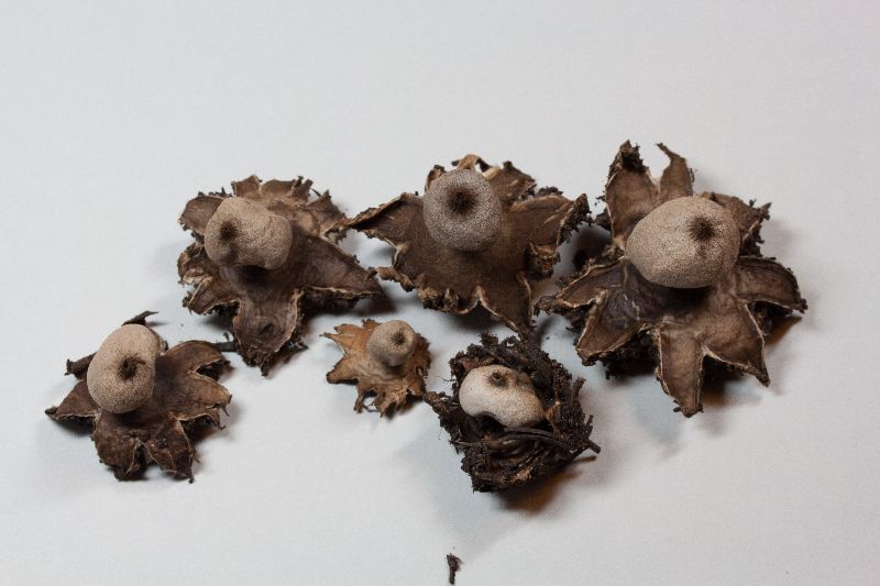
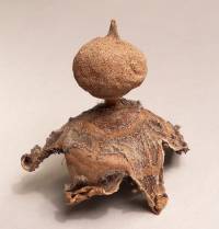
Sunhede S. 1989. Geastraceae (Basidiomycotina): Morphology, Ecology, and Systematics with Special Emphasis on the North European Species. Synopsis Fungorum, 1. Oslo, Norway: Fungiflora Geastrum berkeleyi Massee Ann. Bot. 4:79. 1889 (as Geaster). = Geastrum berkeleyi var. continentale Stanek. Geastraceae. - In Pilat (ed.), Flora CSSR, B1 Gasteromycetes 473-474, 785, 1958. = Geastrum pseudostriatum Hollós, Math. Termesz. Ertes, 19:505-506, 1901 (as Geaster). = Geastrum hollosii Stanek. Geastraceae. - In Pilát (ed.), Flora CSR, B1 Gasteromycetes 467-468, 789-790, 1958. Lectotype of G. berkeleyi (selected here). Collection from Herbarium Mycologicum Berkeleyanum marked: " Geaster multifidus, Lambley, Notts. G. berkeleyanus Mass. and Herbarianum Hookerianum 1867" (herb. K ). Neotype of G. pseudostriatum (selected here). Collection from herb. Hollós marked: "In Robinetis arenosis, prope Kajdaes (Comit Tolna), 21.sept.1927" (herb. BP ). Isotype of G. hollosii. Collection from herb. Stanek marked. "Hurbanovo (Ogyalla, Stara Dala) ap. Nove Zamky, Slovacia merid., Bohemoslovakia, 26.1V.1911, leg. E. Endrey, det. V. J. Stanek. Isotypi" (herb. PRM ). Mature fruitbody mostly small to medium-sized. Mycelial layer encrusted with debris. Exoperidium arched, mostly split into 5-9, nonhygroscopic rays. Endoperidial body rather short-stalked. Endoperidium ± rough. Peristome conical, densely plicate. often ± distinctly delimited. Unexpanded fruitbody: Young fruitbodies, just prior to opening hypogeous, globose to depressed globose. about 1-5.5 cm wide (fig. 24g). Shapes of still younger fruitbodies with whitish gleba shown in fig. 24a,c,d. Mycelial layer encrusted with debris. Expanded fruitbody: Small to large species (tab. 2; fig. 25B). Exoperidium splitting in a star-like way, ± deeply into 4-12, mostly 5-9 rays of varying shape (figs. 23A,B; 25A,B). The exoperidium is ± arched 23A-1:31; 24b,e). In weakly arched specimens the rays are ± horizontally spread (fig. 23A,B) while in strongly arched ones their tips sometimes recurve under the exoperidial disc. Rays not truly hygroscopic but they are sometimes bent or rolled towards the endoperidial body. Diameter of fully expanded specimens 1.5-9 cm (in specimens with horizontally mounted exoperidia 2-13 cm). Pseudoparenehymatous layer when fresh up to 4-5 mm thick, at first whitish, in age turning brownish beige with a reddish tint (cf. Ryman and HolmÀ£sen p. 600. 1984). reddish brown and at last dark to blackish brown, rather easily peeling off or remaining as remnants (in 1-2-year-old fruitbodies often totally disappeared), as dry shrunken and hard, apart from whitish of about the same colours. Fibrous layer as denuded papery coriaceous and dirty white to greyish. Mycelial layer encrusted with debris (fig. 24b), sometimes remaining as a cup in the ground or in age gradually disappearing from the fruitbody. The colour of its inside is brownish beige. Endoperidial body. The endoperidial body is distinctly stalked, often ± globose to very broadly pyriform but varies in shape (figs. 23; 24b,e; 25A; 26). The diameter varies between 6 and 43 mm, mostly 10 and 20 mm (fig. 25B,C; tab. 2). Apophysis often ± prominent or lacking (figs. 26A; 27B:a-c,e,f), sometimes with weak furrows or darker stripes radiating towards the stalk (fig. 26A:j), rarely descending around the stalk as in fig. 26A:k. Stalk relatively short (fig. 26A), (0.5-)1-5 mm high, rounded to oblong in transverse section (fig. 27 B:d), as fresh whitish to light brownish. Endoperidium ± rough, resembling a coarser or finer sandpaper (figs. 23E; 27A). SEM-pictures show processes and short ridges of different kinds (figs. 30A,C-F; 31C,E.F) and crystalline matter (fig. 31B,D). In age the rough surface becomes smoother (fig. 30B). Colour mostly brownish grey to greyish brown but varies (see discussion). Peristome mostly broadly to narrowly conical (acute or blunt), sometimes ± mammiform or rarely almost flat; usually distinctly and densely plicate with 17-36 folds (figs. 23; 24h,i; 26A; 27A:a-f), (< 1-)2-6 mm high; peristome field normally circular to elliptical, mostly distinctly delimited, rarely almost indefinite, often ± deeply depressed or surrounded by a ± pronounced ridge (figs. 23; 27A:a-0, lighter than or concolourous with the endoperidium and without endoperidial processes. A thin mealy peristomal cover of hyphae may be seen in newly expanded specimens. Columella beige to light brown in section, intruding from less to more than the half into the mature gleba, mostly globose to depressed globose or broadly club-shaped (fig. 27B:a-c,e,f). Mature gleba brown. Microscopy: Basidia with basal clamp connection or narrowing into a hyphal part ending with a clamp. as young globose to ellipsoid or ± clavate, as mature of varying shape but often ± ellipsoid with an apical bulge or sublecythiform (fig. 28a-j,m,n.p,q). Mature basidia except hyphal part 12-17 x 6-12 µm; hyphal part <1-14 x 1-2 µm. Pleurobasidia observed (fig. 28k,l,o). Sterigmata 3-7 but mostly 6, 1-2.5 µm long. Subbasidial hyphae thin-walled. 1-2 p.m wide, provided with clamps and ± densely branched. Hyphae of the tramal plates as the subbasidial ones but ± parallel and sparsely branched. Spores in mass brown, as mature, ornamentation included, 5.5-7(- 7.5) p.m in diameter, with an oil drop. light brownish in TRL. SEM pictures show up to 0.8 p.m high. processes (with blunt tips), usually distinct but sometimes ± confluent. The processes are almost equally thick or gradually widening towards the base (figs. 32; cf. 33). The young spore is symmetrically attached to its sterigma, first broadly ovate in age becoming ± globose. Spore wall of fully pigmented spores Cb- and Melz-. Capillitial hyphae 1.5-11.5 µm wide, thick-walled (often with narrow lumen), yellow brown in TRL, Cb- and Melz-, with or without surface debris which may be Cb +. Remnants of thin-walled. clamped hyphae from the young whitish gleba may also be present. Columella hyphae 2-9 p.m wide, thick-walled, pale beige to almost hyaline in TRL. Endoperidial hyphae 2-6 µm wide, thick-walled, crooked (especially in the warts; fig. 29d). Thin-walled, clamped hyphae (of the same kind as of the peristomal cover, see below) have been observed from the surface. Peristomal hyphae 2-10 p.m wide, thick-walled. Hyphae of peristomal cover 2-3 p.m wide (when dilated up to 9 µm wide), thin-walled, clamped, often ± covered with debris. Pseudoparenchymatous layer with thin-walled, bladder-like hyphae of varying sizes (fig. 29c,e). Fibrous layer consisting of 1.5- 8 µm wide, thick-walled hyphae with narrow lumen, almost hyaline in TRL and Cb +. Mycelial layer in the inner part consisting of 1.5- 3(-5) p.m wide, thin-walled, clamped, densely interwoven hyphae (fig. 29b), sparse thick-walled hyphae also present; in the outer part with 1.5-3 µm wide, thick-walled hyphae (fig. 29a).
|
|
|
|


