System maintenance is ongoing as of Monday, October 27, 2025. The portal is currently set to read only with no changes to content available at this time.
Please contact me at help@mycoportal.org if you have any questions.
Agaricus columellatus
|
Agaricus columellatus
 |
|
|
Family: Agaricaceae
[Araneosa columellata Long] ") |
|
") ") The New York Botanical Garden ") The New York Botanical Garden ") The New York Botanical Garden ") The New York Botanical Garden 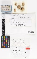 ") ") ") The New York Botanical Garden ") The New York Botanical Garden ") The New York Botanical Garden ") The New York Botanical Garden ") ") |
|
