
|
Pseudobaeospora deckeri C.F. Schwarz
 |
|
|
Family: Tricholomataceae
 Christian Schwarz |
Schwarz CF. 2012. Pseudobaeospora deckeri: A new agaric from central California. Mycotaxon 119: 459-465. Pseudobaeospora deckeri Schwarz sp. nova Latin diagnosis: Pseudobaeosporae pyriferae simillima pileipelle ex hyphis rectis, pileo et stipite KOH coloris alternationem viridis differt. Fibulae praesentes. Basidiosporae 3.1-4.0 x 2.9-3.8 µm, subglobosae vel late ellipsoideae, leviter dextrinoideae. Subter Sequoia. Santa Cruz, USA (California). Holotypus hic designatus: UCSC #7451. Pseudobaeospora deckeri can be distinguished from other members of the genus by its distinctly purple coloration, in combination with alkaline-virescent reactions, a pileipellis which is an irregular cutis with abundant erect elements, lack of a distinctly cellular subpellis, abundant clamps, and lack of cheilocystidia. Fruitbodies collybioid, entirely rubbery-tough to fleshy and slightly cartilaginous when fresh, brittle in age, but rather persistent. Pileus 10-27 mm wide, broadly convex and circular to plano-convex or uplifted and irregularly wavy; margin of young specimens distinctly but narrowly involute and in some specimens distinctly ribbed; very finely pruinose, evenly or in zones or blotches, especially near margin, in age nearly glabrous; deep royal purple (16E-F8) when young and moist, fading to dull brownish or grayish purple (14F4-6), sometimes in age with areas of obscure orange-brown tones (5D5), at margin paler to nearly white. Flesh thin, pallid lilac to darker purple just above lamellae. Lamellae L = 22-38(50), l = 3-5, subdistant to fairly close, adnate to finely and obscurely sinuate, ventricose in age, in some specimens intervenose and transvenose, when young dull gray (8B1-2) to dull lilac (13E3), in age distinctly ochre brownish (6C-D8); margin often thick, even to slightly irregular. Stipe (13-)29-50(-85) x 2-4.5 mm, equal or nearly so, at apex with belts or zones of fine, pale squamules, sometimes quite dense, with a fine pruina below these squamules over the upper quarter of the stipe, especially visible in young specimens, very dark purple (15F8) to royal purple (16D7-8) more or less remaining so at apex, but lower portions becoming duller purple to reddish-brown (11F7, 12D6, 12F4), at base usually conspicuously strigose, this tomentum white to lilac or purplish; base slightly rooting and bound to particles of substrate; flesh solid, dull, grayish purple, cartilaginous. Odor indistinct, although dried specimens wetted with alcohol emitted a ‘mushroomy’ odor (like Agaricus bisporus). Taste weakly but distinctly peppery or acrid. Macrochemical Reactions instantly dark blue-green in 3% KOH, this reaction obscured by the dark color of the fruitbodies and thus best detected by swabbing the treated fruitbody on white paper, or immersing a thin section in KOH. Spore Deposit white. Basidiospores L x W = (2.8)3.6-3.8(4.8) x (2.8)3.3-3.5(3.8) µm. Q = (1.0) 1.05-1.1(1.3). A total of 40 spores from two fruitbodies were examined microscopically: i) (n = 20; from stipe apex, in Melzer’s reagent) 2.8-3.8 x 2.8-4(4.8) µm, Lavg x Wavg = 3.6 x 3.3 µm, Q = 1.0-1.3, Qavg = 1.1. ii) (n=20, from stipe apex, in Melzer’s reagent) 2.9-4.3 x 2.9-3.8 µm, Lavg x Wavg = 3.7 x 3.5 µm, Qavg = 1.05. Globose to subglobose or broadly ellipsoid, some with visible internal droplet; with conspicuous hilar appendage. Morphology and chemical characters of spores variable within a well defined range: immature spores smooth, thin or thick-walled and inconsistently dextrinoid, more mature spores dextrinoid within 5 minutes, thick-walled and smooth. Basidia 24-30 x 3.5-5 µm, clamped at base but obscurely so, tetrasporic, infrequently bisporic, protruding only slightly from lamellar face. Lamellar trama regular, composed of inflated elements 43-80 x 5-28 µm, barrel shaped and tapered at both ends, clamped. Walls irregularly and coarsely thickened. Cystidia absent, but irregularly shaped basidioles at the lamellar margin were inconsistently present. Pileipellis fundamentally a cutis of equal to slightly irregularly inflated hyphae 4.3-8.6 µm in diam., but with many erect elements, thus appearing as a trichoderm, remaining purplish in KOH; erect elements 16-58 x 3.6-6.7 µm, cylindrical to cylindrical-flexuous, often irregularly swollen, constricted or knobbed, often once or twice septate and clamped, often slightly entangled or clustered, more scattered away from disc, not encrusted. Subpellis of tightly interwoven hyphae, not or obscurely differentiated from pileal trama, distinctly green in KOH. Pileus trama parallel, made up of narrow, non-encrusted clamped hyphae. Stipitipellis at apex with a dense coating of clustered caulocystidia over a layer of narrow hyphae 2.8-5 µm in diam., attenuated over upper part of stipe. Caulocystidia 29-40 x 2.9-4.8 µm, cylindrical-flexuous to strangulate or irregularly swollen and constricted or knobby, often onceseptate and clamped; green in KOH (3%). Stipe Trama of inflated hyphae ≤ 8.6-14 µm in diam., pseudoparenchymatous in cross-section, strongly green in KOH. Clamp connections abundant in all tissues. Ecology & distribution - Subcaespitose or scattered, terrestrial. Fruiting at the type locality amongst woodchip mulch in a landscaped area under coast redwood (Sequoia sempervirens). Currently known from three locations: the type locality (University of California Santa Cruz Campus, Santa Cruz Co.), Redwood Camp (Monterey Co.), and Skyline Community College (San Mateo Co.). At the southernmost known locality (Santa Cruz Co.) fruiting near huckleberry (Vaccinium ovatum), red alder (Alnus rubra), and coast redwood, while at the northernmost known locality (San Mateo Co.) fruiting in deep needle duff of Monterey cypress (Hesperocyparis macrocarpa). Fruiting dates include records from early December, January, and March. Additional Collections examined: USA. California: Santa Cruz County, UC Santa Cruz, north side of Engineering 2 Building, 5 Mar 2009, CS 5Mar2009-7 (type mycelium); San mateo County, San Bruno, Skyline Community College, 14 Jan 2010, NS 14Jan2010-3; Monterey County, Big Sur, UC Big Creek Reserve, Redwood Camp, 13 Dec 2010, CS 13Dec2010-5. |
 Christian Schwarz 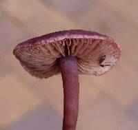 iNaturalist Observations 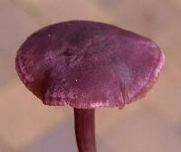 iNaturalist Observations 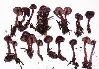 Noah Siegel 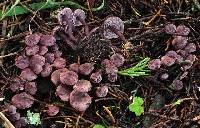 Noah Siegel 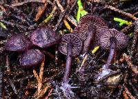 Noah Siegel 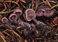 Noah Siegel 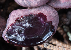 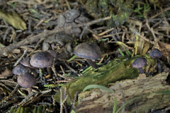 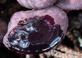 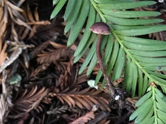 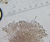 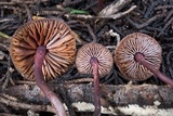 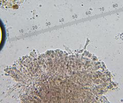 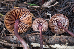 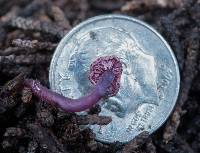 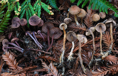 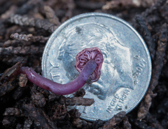 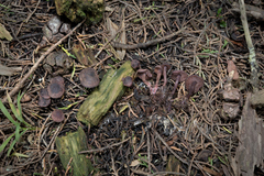 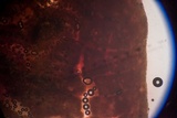 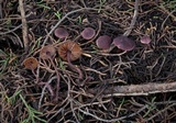  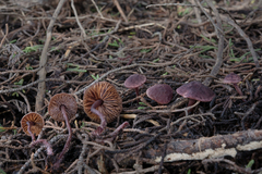 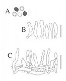 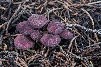 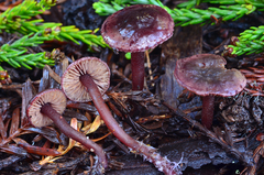 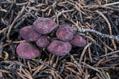 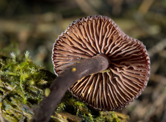 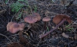 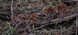 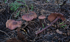 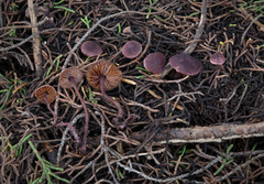  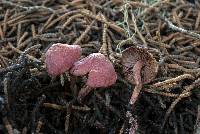 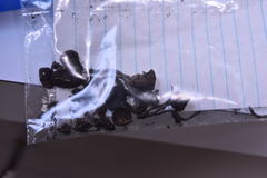 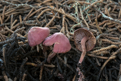 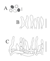 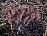  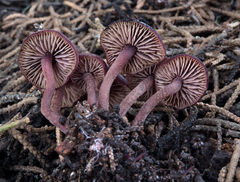  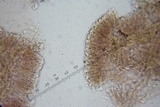 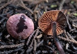 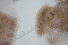 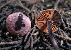 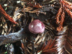 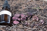 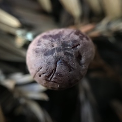 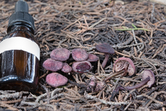 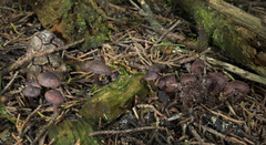  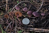  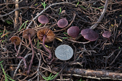  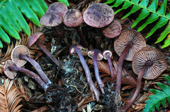 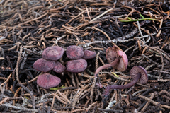 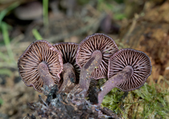 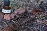 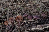 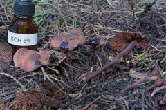 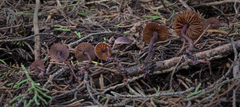 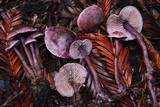 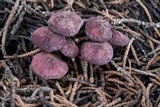 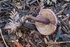 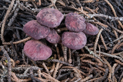 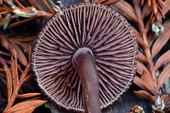  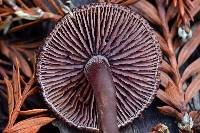 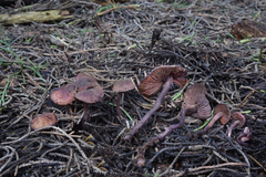 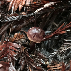  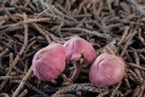  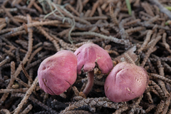 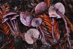 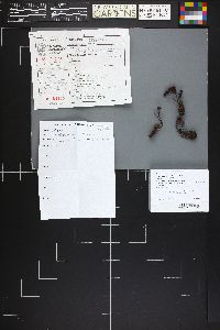 |
|